
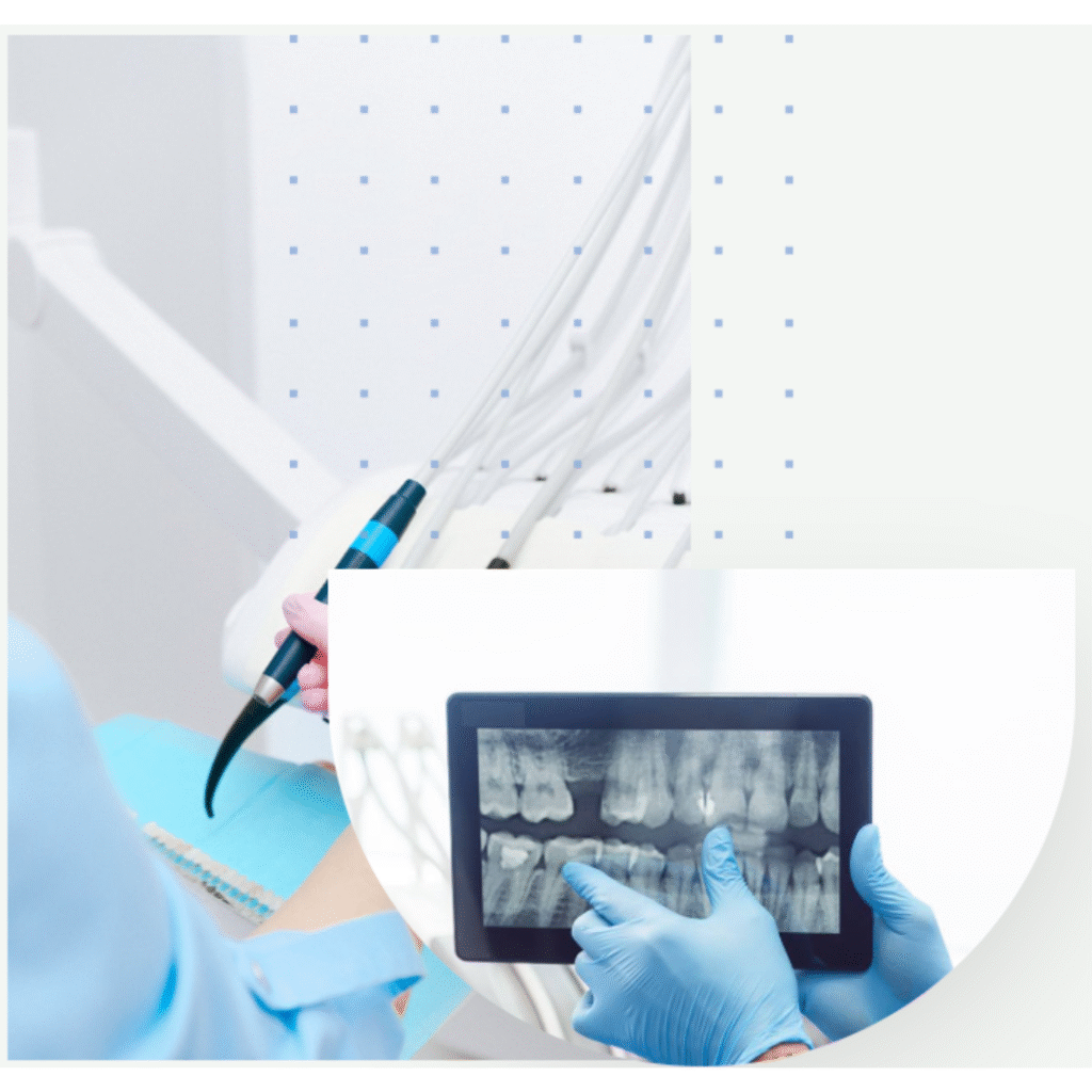
Digital x-rays
Digital X-rays for dental care offer numerous advantages. They provide instant results, reducing wait times. These images are easily stored and shared electronically, enhancing communication among dental professionals. With significantly lower radiation exposure, they prioritize patient safety. Digital X-rays empower dentists for more accurate diagnoses, leading to better oral health outcomes.
FAQs about digital X-Rays
Dental X-rays (radiographs) are internal images of your teeth and jaws. Dentists use X-rays to examine structures they can’t see during a routine checkup, like your jawbone, nerves, sinuses and teeth roots.
Like X-rays taken in other parts of your body, dental X-rays use electromagnetic radiation to capture images of your mouth. The radiation beam passes through your soft tissues and creates images of your teeth and bones.
Dental X-rays may be traditional (taken with film) or digital (taken with digital sensors and a computer). Digital dental X-rays use 80% to 90% less radiation compared to traditional dental X-ray machines.
Dental X-rays
Dental X-rays show cavities, especially small areas of decay between teeth, and decay beneath existing fillings. They help detect bone loss in your jaw and reveal areas of infection. X-rays also show the position of unerupted or impacted teeth, abscessed teeth (infections at the root of your tooth or between your gums and your tooth), as well as cysts and some types of tumors. Dentists use X-rays to determine your eligibility for treatments such as dental implants, braces, or dentures. They also help monitor healing after certain procedures like dental bone grafts and root canal therapy.